The ophthalmoscope
Spot the differences

Checking the eyes
Both vets perform a fundoscopic examination on a dog to check whether its retina and adjoining tissues are healthy. Jakob and Djajadiningrat-Laanen are performing ophthalmoscopy, a method invented in the mid-nineteenth century. Jacob is holding a penlight in his left hand and an eye mirror with a small viewing window in the middle in his right. The mirror reflects the light from the penlight into the patient's eye, right into Jakob's line of sight. The background of the patient's eye reflects the light back, allowing the retina to be examined through the viewing window in the mirror.

Ophthalmoscope 2.0
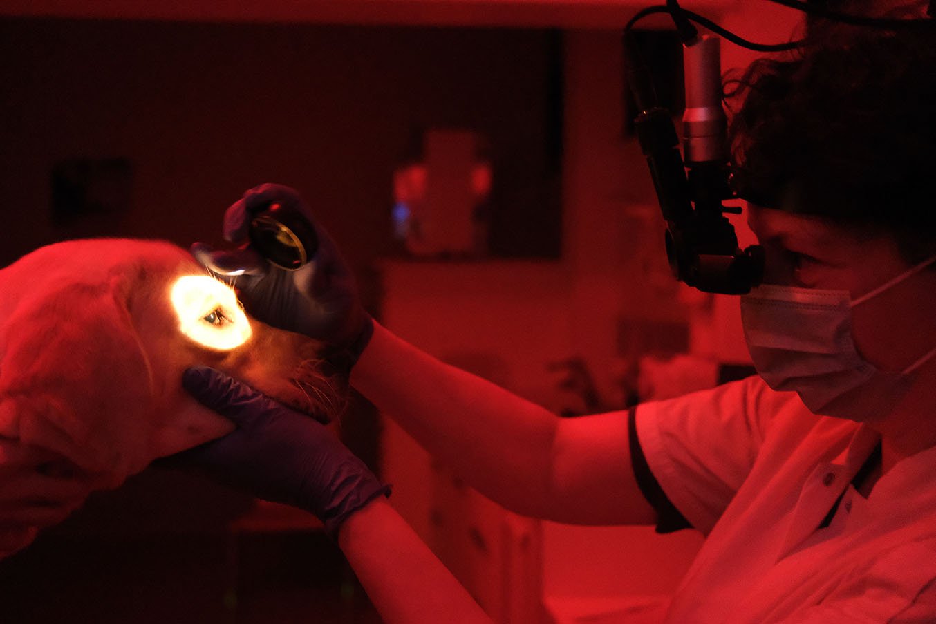
The main difference between the two photographs is the type of ophthalmoscope being used. Jakob is using a direct ophthalmoscope, while Djajadiningrat-Laanen is using an indirect ophthalmoscope that allows the user to observe both eyes (binocular). Binocular indirect ophthalmoscopy has only been in use since the mid-twentieth century. The light is mounted on a headband and illuminates the area the veterinarian is looking at. The veterinarian only needs to hold a lens in order to focus the light on the retina (NB the lens isn't in position yet in this photo). A camera has also been mounted on Djajadiningrat-Laanen's headband, allowing students and patient owners to observe the images in real time on a monitor.
Details in the dark
Other notable differences include the ambient lighting. Jacob and his patient were photographed in a brightly lit room to capture the procedure on film. This photo was originally intended as a teaching aid, allowing students to see how the technique should be carried out on a patient. In practice, the examination actually takes place in the dark as we can see from the more recent photograph. Djajadiningrat-Laanen is working in a darkened eye examination room illuminated only by red light. This makes it easier to observe details.
Short sleeves and a face mask.
Jakob is wearing a white coat with long sleeves while Djajadiningrat-Laanen is wearing short sleeves, in accordance with the prevailing views on hygiene at the time. She is wearing a face mask on account of the COVID-19 pandemic.

