Selective transport in neurons understood
Motor proteins paint the complex road network
Our cells contain a ‘road network’, over which molecules are constantly being transported. In neurons, this transport system is more complex than in our other cells, because nerve cells are not symmetrical and they have long extensions, called axons and dendrites. Molecules therefore have to be selectively transported into right extension. Researchers at Utrecht University have now shown that this selective transport process is made possible thanks to a highly refined, and completely unexpected, organisation of the road network inside neurons. Their findings will be published online in the scientific journal Neuron on 30 November.
The publication is an important milestone in the work of research leader Lukas Kapitein at Utrecht University. It answers the main question posed in his ERC Starting Grant application. The discovery required quite a bit of research, including the development of experimental techniques needed to answer the question. “Using extremely high-resolution microscopy, we were able to take ever-more detailed pictures of the cell, but that didn’t teach us much about how it was exactly organised”, Kapitein explains.
Selectivity vital
Nerve cells have several long extensions protruding from the cell body: multiple dendrites and a single axon. Previous research had indicated that some of the molecules in a nerve cell are only transported into the axon, while others could be transported to both the axon and the dendrites. This selectivity appears to be vital in the transport of molecules. In Alzheimer’s disease, for example, certain proteins can be found in dendrites that should not be there. But how does the cell guide this selective distribution of molecules under normal conditions?
Controlling distribution
So-called ‘motor proteins’ are crucial for arranging transport within the cells. These molecules can move along a ‘road network’ formed by long tubes of proteins, called microtubuli. Each motor protein can only move along the tube in a single direction, either towards the plus end or the minus end. Other research has also shown that some motor proteins can only move along certain types of microtubuli. However, in order to discover how the road network of microtubuli guides motor proteins to the right destination, the researchers in Utrecht required a new technique.

Motor-PAINT
This new technique, called ‘motor-PAINT’, adds coloured motor proteins to a fixed cell. These proteins then ‘paint’ the network of microtubuli that they use to transport molecules throughout the cell, making the precise orientation of the microtubuli visible to the researchers. To everyone’s surprise, Roderick Tas, a PhD candidate working on the project, discovered that microtubuli are not arranged criss-cross among one another, but are rather separated into parallel bundles per type.
Crux
The crux of the transport organisation appears to be the orientation of the various types of microtubuli, and therefore the location of their plus and minus ends. One type of microtubuli is oriented in such a way that it always guides motors to the axon, while others can either lead to an axon or a dendrite. Depending on whether a motor can move over one type or another, it will be guided to the axon or to both axons and dendrites. Thus, the orientation and identity of different microtubules determines which molecules are transported only to the axon, while others can go to either the axon or the dendrites.
Transport system more comprehensible
“Our research shows that the microtubuli in nerve cells are organised in a highly refined and completely unexpected manner. Now that we know about this organisation, the transport system suddenly becomes much more comprehensible. The next challenge is to understand how this organisation is created, and the relationship between the organisation of microtubuli and transport disorders. That will eventually allow us to find out whether transport problems play a role in diseases, and if so, which role they play”, according to Kapitein.

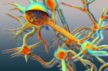
Figure: How the organisation of the road network facilitates both selective and non-selective transport. So-called ‘kinesin motor proteins’ always move with their cargo towards the end of the microtubule marked by a circle. However, the different types of kinesin motor proteins move over different types of microtubuli. In dendrites, these are oriented in opposite directions. Selective transport occurs because different types of kinesin motor proteins binds to different cargoes.
Publication
‘Differentiation between Oppositely Oriented Microtubules Controls Polarized Neuronal Transport’
Roderick P. Tas, Anaël Chazeau, Bas M.C. Cloin, Maaike L.A. Lambers, Casper C. Hoogenraad, Lukas C. Kapitein All authors are affiliated with Utrecht University
Neuron, 30 November, DOI: 10.1016/j.neuron.2017.11.018
This research was funded in part by an ERC Starting Grant awarded to Lukas Kapitein. Further funding was provided by NWO and other ERC grants.
Read more about the research of Lukas Kapitein
- Quantum dots illuminate transport within the cell - publication in Nature Communications
- ‘Road network’ in nerve cell endings visualised in superresolution - publications in Nature Communications
- Researchers control transport in neurons using light - publication in Nature, with video
- A day in the life of a Motor Protein - video
Contact
Monica van der Garde, Faculty of Science, public information officer, m.vandergarde@uu.nl, +31 (0)6 13 66 14 38.
Related news

Quantum dots illuminate transport within the cell
Biophysicists from Utrecht University have developed a strategy for using light-emitting nanocrystals as a marker in living cells.
Cell biologists discover crucial ‘traffic regulator’ in neurons
Cell biologists led by Utrecht University’s Professor Casper Hoogenraad have discovered the protein that may be the crucial traffic regulator for the transport of vital molecules inside nerve cells.