
The new UU Electron Microscopy Centre will be both futuristic and future-proof
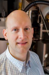
A focused beam of electrons and a very steady sample. These two conditions allow researchers to observe the very smallest of structures using electron microscopy. While the technology has been around for some time, major improvements in the precision of the electron beam have narrowed its focus to a point that is smaller than the space between two atoms. “It’s a pretty exciting idea that we can now observe the molecular structure of complex biomolecules such as proteins, simply by ‘looking’ at them” says Marijn van Huis, head of the centre for Electron Microscopy at Utrecht University. Electron microscopy is pushing the boundaries of what can be made visible. To keep a single molecule in focus, modern electron microscopes need to be incredibly steady. “Someone talking in the next room, a tram passing by or an elevator moving in the building, these things can all influence the measurements ” says van Huis. This is one of the reasons the UU is now building a new centre for electron microscopy.
A much needed new centre for electron microscopy.
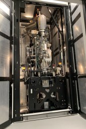
The focus of electron microscopy and that of the research employing the technique is narrowing in tandem. The ability to study structures down to the molecular level has opened up new avenues of research that take the technology to the limits of its capacity. “When we first started with EM it was mostly employed for cell biology. The structures where relatively large for EM so a little vibration was not a big issue,” says van Huis. In their previous location on the 5th floor of the Kruyt building, even the motion of the building had become an issue. “Nowadays you would never consider placing an electron microscope on any floor other than the ground floor,” explains van Huis. The new EM centre only has one floor and the high-end microscopes will be placed on their own concrete foundations, with active anti-vibration to take even the smallest vibrations out of the equation. The machines will be operated from a separate room, to keep researcher interference with the samples to a minimum. “The new building is just far enough away from any tracks and roads to not be influenced by their vibration, but not too far so as not to be visible. The centre will be an iconic part of the Utrecht Science Park, we’ve invested in a top-notch facility.” The microscopes are now housed in a temporary building at the far end of the campus (“right next to the sheep,” laughs van Huis), moving into the new facility will be a big event.
We have around 15 million euros worth of equipment, it would be a waste to have these machines idle for any reason
The new facility will initially house 10 microscopes. There are four spots with active antivibration for the high-end microscopes, one of which is reserved for a yet to be acquired Life-Sciences microscope. The high-end equipment comes with a hefty price tag. An investment of 7 Million euro by the university board in 2016 really took EM in Utrecht to the next level. Additional investment from National Roadmap funds were used to acquire another 3 microscopes. “It’s like we have this fleet of formula 1 cars now. That requires a formula 1 circuit and, most importantly: a team or formula 1 drivers,” says van Huis. “It’s not just the scientists that do important work for the university,” he adds. Highly trained professionals spend years to become expert operators of the EM equipment. “We have around 15 million euros worth of equipment, it would be a waste to have these machines idle for any reason. It all needs to come together in the new EM centre: good housing, good equipment and good people.”

Many researchers in Utrecht involve EM heavily in their research. “In a sense, these are our customers,” says van Huis, who uses only a small portion of the ‘beam-time’ himself. The EM centre is an open platform, meaning outside users can also use it, from external academics to corporate clients, each with their own pricing package. “We have a great facility manager who handles these things,” says van Huis. “The new material science microscope was financed by NWO, it’s the only one of its kind in the Netherlands and serves as a national facility, so at a minimum we have to provide beam-time to external clients for half of its operating hours . With this microscope, we will be the materials science centre for the Netherlands“ says van Huis with a touch of pride.
AntivibrationMuch like noise-canceling headphones, the new platforms on which the EMs will be placed have active antivibration, using piezo crystals that transform with electric currents to stabilise an immense, 3-meter high machine. Humans are the main disturbing factors: we talk, clap, transpire and change the temperature. That’s why the new microscopes are remotely controlled from an operating room, taking away the human element from the experiment. Cancelling electromagnetic fields is much more difficult. There are compensation systems for that but these are much less effective. The best solution is to stay away from any moving metal that might induce a field. |
How does electron microscopy work?
In part, EM works just like a regular, optical microscope. The lenses are not made of glass however but are magnetic. They control the electron beam from the outside. “Electrons are charged particles,” explains van Huis, “and that makes everything different.” With magnetic corrections, the electron beam can be focused to a very narrow point of around 50 picometer (that’s 0,05 nanometer, less than the distance between 2 atoms). The ability to make very fine magnetic adjustments to the electron beam is called aberration correction and the current ‘Thermo Fischer Spectra 300’ microscope is top of the bill when it comes to this. “Using a focused beam to scan across thin materials gives a crystal-clear image even of the atoms,” explains van Huis. This is the first microscope of this type in Utrecht and is part of the national infrastructure NEMI. There are two types of EM: transmission and surface scanning. Transmission looks right through an object (thereby interfering with the sample, which is troublesome in particular for live materials), the other type just scans surfaces and is used by earth and material scientist for instance; both types will be housed in the new centre where everything comes together.
You do not need to be an expert to make good EM pictures, but understanding the physics behind it definitely helps
As a physicist, van Huis graduated on positrons, the ‘anti-particles’ of electrons. Working with bundles of charged particles was not a new thing to him when he developed further to become a nano-scientist, examining structural changes in the smallest of particles in response to external influences such as heat or cold. He performed his first EM experiments to observe these changes first hand. “I am not a life scientist, but as the head of the EM centre I represent both material sciences and life sciences.” As a physicist, van Huis held positions at Delft Technical University and in University of Antwerp. Since 2012, he works at Utrecht University, where he quickly became the head of the EM centre. He still uses the centre for his own research as well, but “With all the administrative duties, less that I’d like to,” he laughs. “Apart from the experiments, in my own research I create an ideal world in a computer simulation that has no external interference. Simulating down to the level of quantum mechanics requires a lot of computing power, especially if you want to model something as complex as a nanoparticle or a protein.” The larger the system, the more compromises you need to make when modelling it. Using EM to feed computer simulations and verify the outcome of such simulations requires knowledge at both ends. “That’s also the beauty of physics: take disinfectants for example, they work at the molecular level by adding electrical potentials to proteins thereby disabling bacteria. To understand these concepts, you need both biochemistry and physics. The same goes for protein folding for instance, which is a time-dependent process that is hard to simulate. Researchers such as Professor Alexandre Bonvin at the Bijvoet institute are very proficient in that area of research.
The professor in cryo-EM Friedrich Förster has a background in Physics and Biophysics, and Professor Lukas Kaptein in Biology also has a physics background. “It helps with the interpretation and when you’re making sensitive recordings.”. It’s not just a matter of getting a pretty picture, understanding the image formation mechanisms and how to use the electron beam as effectively as possible are of paramount importance. Using a beam of electrons damages or destroys small biological structures, so you need to use tricks to keep the electron dosage and interference at the lowest possible level. The argument also goes the other way: physicists lack the knowledge in life sciences such as cell biology or protein science. That is why collaboration is key (and Utrecht is very good at that!). People skilled in both areas are ideal, but the more you move towards chemistry and biology, the more empirical knowledge becomes important. “Biological systems are incredibly complex and heterogenous. You can’t just change a single variable and measure effects separately, it’s often more than 20 things that happen simultaneously. That would drive a physicist mad,” exclaims van Huis.
Life Sciences and EM: minimal damage to living material

Life scientists are often interested in a specific protein or other biological structure. To get a clear EM picture, there are roughly two routes: either you isolate the desired structure (hard, but not impossible to do) and study that one ‘object’ in great detail under EM, or you can use purification to obtain a lot of proteins of the same type and you make many different pictures of many different copies of the object and use computers to reconstruct the structure. Another option is to freeze a particular microbiological scene (a protein in action, for instance) and then carefully rotate the sample under the EM (tomography). Rotating a biological sample from -90 to +90 degrees and taking pictures roughly at every degree can be used to extrapolate the 3D structure. Typically, you rotate by 1 degree, correct for x-y shift and re-focus, take another picture. Focus and position are determined by software. To minimise exposure to the electron beam, you can go faster, forego the focus correction or do this live using predictive algorithms, proteins are sensitive to radiation so shorter time is better! In the future we can do these corrections automatically using artificial intelligence (AI). Researchers are already on this field, developing AI-like optimisation algorithms. Also, the reconstruction itself (not only in electron tomography, but also in MRI and MircoCT (Cat scans in hospitals in hospitals) profits from AI. Artificial Intelligence is not the answer to everything though, it can make bad predictions and always needs verification.
As systems get better at predicting, the software needs less and less images to extrapolate structure, so less destruction. But there is a catch in that when used exceedingly, one ends up with manipulated data. You always need to re-check and calculate, and offset the AI result to an actual old-fashioned measurement. For large-scale commercial purposes that require huge amounts of data to be processed (such as in semiconductor industry), AI can save time. Also it can be used to interpret the images obtained by EM. Many EM recordings of cells, for instance, are still manually coloured in, often in huge centres with hundreds of people colouring cell-organelles. Presently, algorithms are not good enough for that, but it is our hope that AI can solve this!
Future outlook
Utrecht University and UMC Utrecht are jointly known as ‘Electron Microscopy Utrecht’. The new EM centre will only house the UU microscopes. “We’d welcome the UMC Utrecht microscopes in our facility, but that is administratively difficult,” says van Huis. "UMC Utrecht is well known for its cell biology and correlative microscopy (a combination of optical and electron microscopy), so when we have research question that require it we can easily go to their center. In turn, we are part of the NEMI (Netherlands Electron Microscopy Infrastructure).There is a whole community around microscopy. Our facility manager does a wonderful job in matching equipment to end-users. Beam-time can also be requested through the NEMI
Every scientific discovery that uses EM is a success for us. We can achieve more if disciplines collaborate, which is one of the strengths of Utrecht Life Sciences. When we can bring top experts together in the fields of catalysis, viruses, proteins etc. and combine that with our expertise in physics, we will be a winning team. Our EM centre is already internationally well-known for its contributions to global challenges in improving EM technology. In Utrecht, we are very good at in situ observations, building miniature experiments that can be run within the microscope to show live biological processes and transformations in nanoparticles. Technology such as biology on a chip can fit into our machine, it needs to be very small, though (10cm x 1cm x 1mm), even keeping biological systems alive until we switch on the electron beam, which shows us a brief glimpse of life at the molecular level. And that’s exciting stuff.