Structure of protein connecting neuron and myelin clarified
Nature Communications publication
Researchers from Utrecht have resolved the structure of a protein that is responsible for the connection between the neurons and the cells that form the protective layer around them. This protein, called MAG, is also responsible for the exchange of information between the cells. As MAG is involved in the recovery of neurons, this knowledge may be useful in the search for treatments for diseases in which the nervous system is damaged. The researchers published their findings in Nature Communications on 6 December 2016.
In order to ensure that nerves can transmit their signals fast enough through our nervous system, they are enwrapped by a fatty substance called myelin. The protein myelin-associated glycoprotein (MAG) plays a major role in the formation and maintenance of the myelin. It is located between the cell membrane of the neuron and that of the cells that form the myelin. When neurons are damaged, for example due to spinal cord injury or a stroke, MAG actively inhibits recovery.
“If you could switch off the protein, you might be able to stimulate the regeneration of the neuron”, says Bert Janssen, chemist at the Department of Crystal and Structural Chemistry and leader of the research. “That’s why we are so interested in the exact structure of MAG.”
If you could switch off the protein, you might be able to stimulate the regeneration of the neuron.
Three-dimensional structure
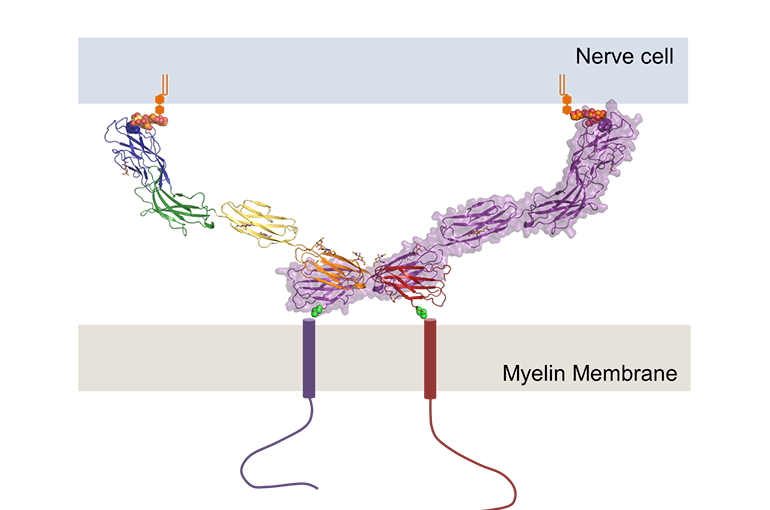
PhD student Matti Pronker and research supervisor Bert Janssen studied the MAG protein crystals using X-ray diffraction. This allowed them to uncover the three-dimensional structure of the part that bridges the two cell membranes, from which they were able to derive how MAG recognises and binds to sugar groups on the surface of the neuron.

They also discovered that a MAG molecule can link with a second MAG molecule to form a dimer complex. This link occurs on the cell membrane of the cell that forms the myelin. With the dimer formation on one side of the molecule and the interaction with the sugar molecules on the other, MAG is able to regulate the distance between the two cells. “It’s like a rigid stick between the two cells, holding them at a distance of around 10 nanometres”, explains Bert Janssen, who conducted the study with the help of the Vidi grant he received from NWO in 2012.
Signal exchange
The chemists also illustrated how dimer formation is important for the communication between the neuron and the myelin-forming cell. Janssen: “The dimer penetrates the membrane of the myelin-forming cell, allowing it to convey messages to the cell.”
In collaboration with researchers at UMC Utrecht, they were able to prove that both dimer formation and the interaction with the sugar molecules are important for neuron growth.
Publication
Matti F. Pronker, Suzanne Lemstra, Joost Snijder, Albert J.R. Heck, Dominique M.E. Thies-Weesie, R. Jeroen Pasterkamp and Bert J.C. Janssen.
Structural basis of myelin-associated glycoprotein adhesion and signalling.
Nature Communications, 6 Dec 2016. Doi: 10.1038/NCOMMS13584