New Utrecht-based lab to focus on brain development
Utrecht University and UMC Utrecht open MIND Facility
On Friday 2 June, Utrecht University and UMC Utrecht opened the MIND Facility, an innovative research centre clustering the world's foremost expertise in the field of neurological research. Here, the most advanced stem cell technology and microscopy will open the way to a better understanding of the origins and development of the human brain. And also of what can go wrong, why, and how science can intervene.
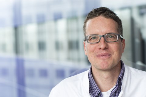
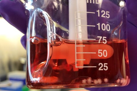
In the MIND Facility, researchers will be able to study the brain using pieces of 3D brain tissue grown from human stem cells, known as organoids. 'Using lab-based techniques we can derive stem cells from ordinary hair, blood or skin cells, which can then be developed into all kinds of tissues by placing the stem cells in conditions resembling the tissue's natural environment', explains Jeroen Pasterkamp, professor of Translational Neurosciences and director of the MIND Facility. 'We mimic in the lab what takes place in the brain during embryonic development. The cells then start building 3D brain tissue, enabling us to study the development.'

Though these tissues comprise only sections of the brain and in no way replicate the real thing, 'it is a fantastic tool for answering all kinds of questions about how the brain and brain cells function', says Pasterkamp. 'You can see neurons' electrical activity and see them making connections. Everything you see in a real human brain, we can see in the lab, too.'
The MIND Facility aspires to attract researchers from a wide range of disciplines. Pasterkamp: 'Working together, we'll be able to unravel how the immature brain develops, and in the case of disorders discover what goes wrong. To do that, we want to reproduce and study brain areas in the lab that are important for normal childhood development, as well as areas that are involved in developmental and other disorders.'

By comparing the stem cells of healthy children and children with a developmental disorder, scientists will be able to pinpoint at a cellular level where aberrations arise. 'We can genetically modify stem cells before they begin forming into tissue, enabling us to introduce a mutation that is common among people with autism spectrum disorder, for example. Then you can compare the mutated organoids with tissue that doesn't contain the mutation and ask: what are the differences?'
Everything you see in a real human brain, we can see in the lab, too.

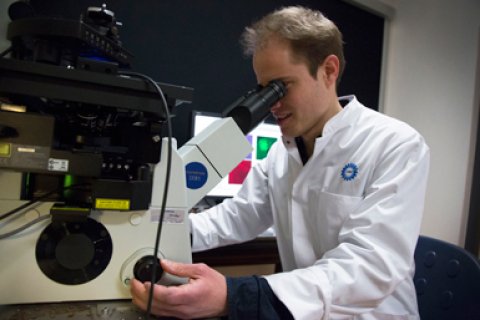
Unlike two-dimensional tissue samples, organoids cannot be placed on a Petri dish and studied under a microscope. 'We’re talking about a clump of cells. UU and UMC Utrecht have therefore invested in an innovative technology called light sheet microscopy', Pasterkamp continues. 'If you make the tissue transparent, you can use this technique to render it into digital cross-sections, which then creates a 3D image of the brain tissue. This allows us to see the precise composition of sections of the brain, including which types of cells are found there.'
The ambition of the MIND Facility is to become one of the world's leading brain organoid labs. 'We have two light sheet microscopes that are available to anyone interested in conducting scientific research experiments on human brain tissue.' In the future, organoids may partly replace the use of animal models in the lab.
MIND (Multidisciplinary Investigation of Neural Disorders) is an alliance of the Brain Center Rudolf Magnus and the Faculty of Science at Utrecht University.
Within Utrecht University’s strategic research theme Dynamics of Youth, Jeroen Pasterkamp is now developing a new interdisciplinary research project on early brain development: The first 1001 critical days of a child’s life.

