
New insight into brain development disorder microcephaly
Publication in Nature Cell Biology:
Two years ago, the Zika virus drew attention to microcephaly, a developmental disorder in which the brain and skull display inhibited growth. But there are other causes of microcephaly, such as congenital genetic diseases. Much is still unknown about brain development, but researchers at Utrecht University, in collaboration with their colleagues in Switzerland, have now new shed light on the molecules involved. The results of their research are published in Nature Cell Biology of 24 April.
“Biological processes are determined by molecules in our cells. We can only understand the factors that determine health and disease and find medicines to control these factors by zooming into this molecular world”, explains research leader Prof. Anna Akhmanova.
Surprising discovery
The researchers began their studies by focusing on the protein ASPM. “We knew that the genetic form of microcephaly is most often caused by defects in this protein. But a surprising discovery was that ASPM appears to work closely together with another protein, called katanin”, tells Akhmanova.
When the balance sways too much in one direction or the other, too few brain cells are produced
Essential for healthy development
It appears that precisely this collaboration is important for cell division, and therefore for the normal development of brain cells. “The interaction between ASPM and katanin is required for the proper balance between cell division and their specialisation into nerve cells. When the balance sways too much in one direction or the other, too few brain cells are produced”, Akhmanova adds.
Crucial balance
For developing brain cells, this balance is especially crucial, because once they become nerve cells, they cannot divide. If new cells develop into nerve cells too quickly, not enough cells are formed, and the brain remains small.

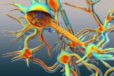
Key role
During cell division, DNA must be copied and distributed between daughter cells. A cellular structure called the mitotic spindle pulls apart the DNA-containing structures, the chromosomes. The photo shows a microscopic image of DNA (blue) in a spindle. The protein ASPM appears to play a key role in this process, as it is located at the ‘poles’ (yellow) in the spindle.
Spindle position
The study shows how ASPM does its work at the molecular level, and why it is so important. In cooperation with the protein katanin, ASPM is responsible for the regulation of the organisation and positioning of the spindle. “It is this positioning that helps to determine how the daughter cells develop: will they become copies of new cells, or will they develop into nerve cells”, Akhmanova explains.
Much broader insight
The fact that a deviation in the protein ASPM leads to microcephaly can now be better understood at the molecular level. However, the results of this study provide a much broader insight, which may make it possible to explain or find other causes of the disorder.
Evolutionarily unique
Akhmanova’s fascination for brain development is not limited to disease, however. “Even apes, our closest relatives, have much less brain capacity than we do. Our brain makes us what we are. This means the development of our brain is evolutionarily very special.”
Publication
“Microtubule minus-end regulation at spindle poles by an ASPM–katanin complex”
Kai Jiang*, Lenka Rezabkova, Shasha Hua*, Qingyang Liu*, Guido Capitani, A. F. Maarten Altelaar*, Albert J. R. Heck*, Richard A. Kammerer, Michel O. Steinmetz and Anna Akhmanova*
Nature Cell Biology 24 April 2017, http://dx.doi.org/10.1038/ncb3511
* Utrecht University
More information
- Website Cell Biology
- This research is part of Science for Life, one of the four themes of Utrecht University’s interdisciplinary research programme Life Sciences.
Contact
Monica van der Garde, Public Information Officer, m.vandergarde@uu.nl, +31 (0)6 13 66 14 38
Related news
Cell biologists discover crucial ‘traffic regulator’ in neurons
Cell biologists led by Utrecht University’s Professor Casper Hoogenraad have discovered the protein that may be the crucial traffic regulator for the transport of vital molecules inside nerve cells.

Protein found that determines critical step in brain development
Cell biologists at Utrecht University have discovered a key protein that serves as the basic element for the intracellular structure of nerve cells.